The Hoof and Its Relation to Balance and Soundness
The association between man and horse is a long and storied one. The relationship has endured the use of the horse by man as a source of food, an ally in battle, a partner in the working world and a source of enjoyment on the racetrack, in the show ring and on the trail. Throughout this long association, man has forever altered horses as a group and as individuals through selective breeding, training and cosmetic adaptations. From the size and shape of a horse’s ear to the length and thickness of his tail, man has been responsible for a great many alterations.
Evidence of genetic adaptations and man’s manipulative influence can be seen in the structure of the equine foot. Throughout the evolution of the horse the hoof has changed to adapt to its environment. Horses developed attributes that allowed them to better survive in the types of areas where they existed. Horses in wet, marshy areas developed large, flat hooves that enabled them greater purchase in the soil. Plains-dwelling horses developed small, hard hooves that provided them swift flight across the solid ground.
Man’s selective breeding practices saw further adaptations. The Clydesdale, among other attributes, was bred over time to have large, durable feet that were deemed necessary to pull heavily laden wagons across hard surfaces. The larger hooves dispersed the shock of impact on the firm ground and helped the animal stay sound.
Selection has adversely affected hoof integrity in certain breeds. Recently, in the Quarter Horse breed, it was considered desirable for the horses to have small delicate hooves. In selecting for small feet on heavily muscled, large boned animals like the Quarter Horse, the occurrence of navicular disease has skyrocketed.
Structure
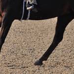
Though the size of the hoof may differ from breed to breed, the basic structure and shape are the same. Left to nature’s influence a horse’s hoof will tend to be rounded or slightly oval. The hoof contains an outer wall, the sole, the frog, bones, cartilage, tendons and a blood supply that all play very important roles to support the weight of the entire animal.
The outer wall of the hoof is made up of keratinized epithelial cells that run parallel to each other from the coronary band to the ground. These cells give the hoof its hard, horny surface. The sole of the hoof is comprised of similar material to the hoof wall. The sole is a layered structure and slightly less porous than the outer wall. The sole is not meant to be a primary weight-bearing surface but helps to protect the sensitive tissues beneath it and serves to hold the outer surfaces of the wall apart. It also serves as a support structure for the wall. The frog is a soft, elastic v-shaped shock absorber that should always have contact with the ground when the foot is bearing weight. The digital cushion lies at the back half of the foot and acts as another shock absorber.
Five corium’s (nutrition-bearing vascular tissues) are essential to the growth and health of the hoof and are located in and at the coronary band, the laminae, the sole and the frog. Inside the hoof wall and the sole is a delicate support structure made up of bones, cartilage and tendons that all depend upon the healthy exterior of the hoof for support and protection. There are three bones located in the hoof.
The short pastern or second phalanx is partly in the hoof and partly above it. The coffin bone or third phalanx is a porous large bone and is almost located entirely within the front of the hoof. The navicular bone is the smallest of the bones and serves to help in the movement of the coffin joint. The lateral cartilages are attached to the coffin bone and rise above the coronary band. They help to compress the veins that drain the hoof. The deep digital flexor tendon originates on portions of the humerus, radius and ulna. It inserts on the third phalanx (coffin bone). The superficial digital flexor tendon originates on the humerus and radius. This tendon is attached to the first phalanx (long pastern bone) and second phalanx (short pastern bone) behind the collateral ligaments of both structures.
This delicate arrangement provides the horse with the ability to move freely and prevents the leg from hyperflexing. The primary blood supply to the hoof comes down the back of the leg on either side of the tendons and goes through small holes in the coffin bone to supply oxygenated arterial blood to the hoof structure.
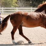
The hoof can be divided into three sections. The heel at the rear of the hoof contains the cleft of the frog and the bulbs. The quarter is the area between the heel and the toe. The point of the frog is in this area as well. The toe is the front of the hoof where the wall and sole are often thickest. Whenever a horse is moving, regardless of the gait, the frog acts as a shock absorber as the horse begins to put the full pressure of his weight upon the foot. The foot should then roll forward to the point where, as it leaves the ground, the toe bears the weight as the horse pushes off with that hoof, breaking squarely over the toe.
Balance
For a horse to be able to move in a balanced fashion, it must have certain attributes. Looking at the horse from the side, an imaginary line dividing the front leg into two equal parts should be able to be drawn from the spinous process of the shoulder blade down through the center of the leg to a point just behind the heel. On the back leg, a line should be able to be drawn from the point of the pelvis to touch the point of the hock, run down the rear aspect of the cannon and touch the ground about three to five inches behind the heel. The hoof wall should slope at the same angle as the pastern, which in turn should mirror the slope of the shoulder. A line following the slope of the rear coronet should be able to be drawn from the ground through the hoof to meet a point within an inch of the back of the knee on the front leg. For the average horse the correct angle for the hoof will be in the neighborhood of 45° to 55° which will allow for the stride to be a symmetrical arc from the point of takeoff to landing. Not every horse possesses perfect conformation
Depending upon the problem, the end result to the animal can be catastrophic or relatively benign. A horse with a club foot will have a different natural arc to his stride which will put different stresses on the bones and muscle support system. A horse with a longer toe will typically have a longer stride, but the toe will hold the hoof on the ground longer putting unnatural stresses on the bone and supporting structures. Long toes can cause a strain on flexor tendons, the suspensory ligament, and the proximal sesamoid bones. Inappropriate angles change the flight of the hoof, which affects the symmetry of the body. A short toe with a high heel equals increased concussion to the hoof, thus increasing the incidence of navicular disease, ringbone, and arthritis of the fetlock. If the angle is allowed to remain incorrect, changes occur in the supporting structures as the body attempts to compensate. This compensation can lead to serious discomfort and lameness.
Flat-footed horses are very sensitive to the type of footing on which they live and exercise. They are intolerant of exercise on rough ground and may require pads to help them to be comfortable. Careful attention to providing corrective trimming and shoeing can greatly help horses that have hoof abnormalities.
Most Popular
 Putting Weight on a Skinny Horse (327,006)
 Benefits of Beet Pulp for Horses (217,487)
 Hot Blood, Warm Blood, Cold Blood in Horses (174,514)
 Possible Link Between Selenium and Cribbing in Horses (163,867)