Laminitis Basics
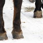
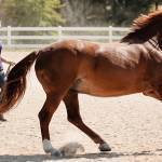
The stance is unmistakable: forelimbs positioned oddly in front of the chest and hind limbs planted under the abdomen in a desperate attempt to support the heft of the horse. This classical “sawhorse” posture is undeniably indicative of the insidious syndrome known as laminitis. Succinctly put, laminitis is the local manifestation of a body-wide metabolic breakdown. Why this metabolic collapse materializes in the feet is not completely understood, but the damage that occurs is well documented.
The Anatomy of Laminitis
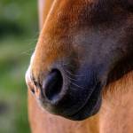
Of the two bones that are completely encapsulated in the hoof, the coffin bone (also known as the distal phalanx or pedal bone) is the largest and is shaped much like the hoof. The significantly smaller, shuttle-shaped navicular bone lies adjacent to the coffin bone and closer to the heel.
Nestled between the coffin bone and the hoof wall are a series of interlocking tissues called laminae. Sensitive laminae are produced by the corium or outer layer of the coffin bone and mesh steadfastly with insensitive laminae, which line the hoof wall, to create a delicate, accordion-like structure that supports the coffin bone. The folds of the laminae are expansive, with some researchers calculating the surface area of the laminae to be approximately that of a tennis court when unfurled.
One of the most critical functions of the laminae, both the sensitive and insensitive, is to anchor the coffin bone against the pull of the deep digital flexor tendon, one of the largest tendons in the forelimb. When this mainstay of support is jeopardized, no matter how slightly, laminitis may result.
How It Happens, In a Nutshell
Laminitis occurs when the cells of the laminae do not receive sufficient nutrients from the blood supply.
Undernourishment sets the stage for inflammation and eventual cell death. Due to a mechanism not well understood by researchers, blood is actually shunted away from the foot, which accounts for the oft-found bounding pulse of laminitic horses. As the laminae become damaged, they weaken and are unable to keep the coffin bone in place, and in severe cases, the tip of the coffin bone shifts toward the sole of the hoof. Complete destruction of laminae can occur within hours of the initiating insult. Once crumbling of foot architecture commences, the horse begins to experience pain that may in time be excruciating and eventually career-ending. Laminitis most often affects both front feet simultaneously. When only one forefoot is affected, it is usually caused by excessive load-bearing due to injury of the opposite leg. Much less frequent is the occurrence of laminitis in the hind feet without involvement of the front feet.
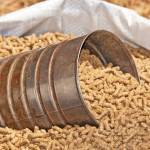
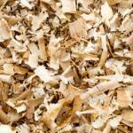
Gorging on starch-rich grains. Standing knee-deep in black walnut bedding. Drinking copiously following sweat drenching exercise. Grazing unlimited amounts of lush pasture. These factors and several others are well-known causes of laminitis. Two relatively new causes of laminitis that have come to the forefront of recent research are ingestion of fructans and obesity.
The Fructan Factor
Well-tended pasture can fulfill the nutritional requirements of many horses and is therefore often the foundation of sound feeding programs. Several species of sugar-rich grasses are planted in hopes of increasing production in cattle and sheep. Little thought is given to the effects these grasses may have on horses. These temperate grasses frequently contain large amounts of water-soluble carbohydrates such as sucrose, fructose, and glucose. In addition, these plants are rich in fructans, which are simple chains of fructose molecules.
Researchers believe that fructans cannot be digested in the stomach or small intestine of the horse. They can, however, be utilized by certain microbial species in the hindgut but only at the expense of the entire intestinal ecosystem. Researchers theorize that when large quantities of fructan gain entrance into the hindgut, a rapid change occurs in the microorganism population, including the death of vast numbers of bacteria. As these bacteria die, they release endotoxins which pass easily though the wall of the intestine and enter the bloodstream. For reasons that are not entirely clear, this endotoxin insult instigates founder. This is the same mechanism that triggers laminitis following the over consumption of grain.
Researchers at the Institute of Grassland and Environmental Research in Aberystwyth, United Kingdom have completed extensive research involving the fructan concentrations of temperate grasses popular in that country.
In their study, the researchers discovered that time of grazing was critical in determining fructan ingestion. Using a single pasture, it was found that horses grazing from 9:00 a.m.to 3:00 p.m.could consume approximately 2 kg of fructan. Conversely, those grazing from 3:00 p.m.to 9:00 p.m.could potentially ingest only 500 g or one-fourth of that consumed during midday grazing. From this data, researchers concluded that the fructan content of grasses changes dramatically, not only day to day but also within a single day. Fructan concentration is affected by exposure to sunlight, temperature, water availability, grass species, and a host of other lesser factors.
In another experiment by the same researchers, horses grazing certain pastures could devour approximately 5 kg of fructan in one day, which is similar to the amount of cereal starch known to induce laminitis in horses. Christopher Pollitt, BVSc, PhD of the Australian Equine Laminitis Research Unit at the University of Queensland, has tested the fructan theory in an experimental setting. In preliminary trials, he and his coworkers brought about laminitis using commercially available fructan. The amount of starch in the dose was only about half of that formerly proven to induce laminitis. Signs of mild laminitis occurred in dosed horses in 48 hours.
Scientists continue to work fervently on the fructan theory, primarily because of the considerable number of unanswered questions surrounding it. Pollitt hopes to answer some of these questions using data from an ongoing trial. The goals of the present study are formidable: to invent ways to reduce fructan in pastures, to pinpoint methods of limiting fructan consumption, and to stabilize hindgut bacteria when confronted with fructan to prevent the formation of laminitis triggers such as endotoxins.
Until more conclusive management techniques are forwarded from researchers, there are ways to keep laminitis-prone horses out of harm’s way. These horses should only be allowed to graze pastures with grass species known to produce small quantities of fructan. Regulating time of grazing is another management strategy. By avoiding pasture exposure during the midday hours, horses are unlikely to consume sufficient fructan to cause laminitis.
Obesity-Associated Laminitis
Obesity-associated laminitis is frequently observed in adult horses that do not receive consistent work. Those affected with this syndrome typically have distinctive body fat distribution, with large pockets of fat deposited on the crest of the neck and the rump. Male horses also tend to accumulate fat in their sheaths. Horses prone to obesity associated laminitis are often difficult to manage nutritionally, as reducing calorie intake does little to lower the body condition of these horses. This syndrome may be particularly frustrating on breeding farms. Affected broodmares may experience irregular estrous cycles, which in turn lowers reproductive efficiency.
Obesity-related laminitis is often incorrectly linked to hypothyroidism. This mistake is made most frequently because some affected horses have low serum thyroid hormone concentrations. Scientific studies, however, have proven conclusively that this specific combination of obesity and laminitis is unrelated to disordered thyroid metabolism. Thyroid stimulation tests have not elicited expected abnormal responses in these horses, as they would have if the horses were suffering from the primary differential diagnosis, Cushing’s syndrome. In addition, experimental removal of the thyroid glands did not induce obesity or laminitis but did produce other signs indicative of true hypothyroidism. Therefore, the potential role of hypothyroidism in obesity-associated laminitis is questionable.
Once Cushing’s syndrome has been ruled out as a possible cause of the laminitis, veterinarians should look to the obesity-associated syndrome. Philip Johnson, BVSc (Hons), DVM, Dipl. ACVIM, MRCVS, associate professor in equine internal medicine at the University of Missouri, believes the abnormal activity of a single enzyme may be the root of obesity-associated laminitis. This enzyme, named 11- beta hydroxysteroid dehydrogenase (HSD), is responsible for cortisol regulation within cells.
The versatility of HSD is remarkable. Based on cellular environment and requirement, the enzyme is capable of converting cortisol to cortisone, its inactive metabolite, and when necessary, the enzyme can change cortisone back to cortisol. This enzyme efficiently dictates cortisol concentration of the cell independent of circulating levels.
Johnson developed a reliable test to ascertain HSD levels in peripheral tissues such as skin, fat, and laminar tissue. Once he found that HSD concentrations could be measured in skin and laminar membranes, he compared levels of HSD in healthy adult horses to those found in laminitic horses. HSD was noticeably elevated in tissues obtained from horses with laminitis. Additionally, the form of HSD responsible for converting inactive cortisone to active cortisol was markedly elevated. Results of this study suggest that hyperproduction of cortisol by peripheral tissues could be a factor in the clinical appearance of obesity-associated laminitis.
Recent studies in human medicine have revealed increased cortisol levels in cells caused by disturbed HSD activity. The result of this enzyme disturbance was the appearance of symptoms similar to those of Cushing’s disease. Elevated HSD activity in cells leads to increased cortisol in tissues. This accumulation of cortisol is an important cause of a syndrome in humans called central obesity or omental Cushing’s syndrome.
Humans with central obesity and horses with obesity associated laminitis have significant similarities including abnormal distribution of fat stores, difficulty losing weight, and reduced fertility. Horses and humans affected with the disease tend also to have hypertension, mild hyperlipidemia, and insulin resistance. The relationship between the species-specific syndromes is obvious, and the human disorder continues to serve as a model for equine researchers.
While obesity-associated laminitis is not well understood among researchers and veterinarians, affected horses may go on to lead otherwise healthy lives if treatment is swift and diligent. Recommended treatments center around corrective trimming and shoeing, use of nonsteroidal anti-inflammatory medications for pain, and strict diet. Forced exercise can be imposed once all laminitis-related pain has abated.
Prognosis
The prognosis for horses faced with complicated laminitis is not bright. The majority of horses with laminitis recover using various means of treatment—some even without treatment—and seem to suffer no ill consequences. Nearly 20-30% of affected horses will, however, have devastating outcomes resulting in catastrophic damage to the laminae. If these horses are able to survive, the likelihood that they will return to work is tenuous. Laminitis is unquestionably a devastating disease. As researchers delve deeper into the causes of this disease, the likelihood that horse owners will one day be able to prevent its occurrence is promising.
Most Popular
 Putting Weight on a Skinny Horse (326,683)
 Benefits of Beet Pulp for Horses (217,223)
 Hot Blood, Warm Blood, Cold Blood in Horses (174,383)
 Possible Link Between Selenium and Cribbing in Horses (163,775)